Diagnostic Ultrasound Machines help clinicians examine soft tissues and moving structures without using ionizing radiation. In a busy clinic, a clear image can support a timely assessment of the abdomen, heart, or developing pregnancy. The technology is widely used, but its value depends on more than the screen. Probe selection, image settings, patient position, and operator experience all influence what appears.
A well-chosen system can fit the realities of a practice: patient volume, available space, specialty, and staff training. A portable unit may help during bedside examinations, while a cart-based system can offer a broader set of functions. Neither option is automatically better. The right choice should match the examinations performed most often and the level of detail clinicians need. Small controls matter. So does a readable display under bright room lights.
Reliable purchasing also means looking beyond advertised features. Buyers should review image quality in relevant applications, probe availability, service support, software updates, and the cost of maintenance. Demonstrations with representative cases can reveal workflow strengths—and limitations that a brochure may not show. No machine replaces clinical judgment, and ultrasound findings may require confirmation through other methods. That deserves honest consideration. Comparing Diagnostic Ultrasound Machines carefully helps healthcare teams choose equipment that supports consistent examinations, practical workflows, and informed patient care.
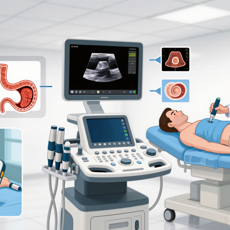
What Diagnostic Ultrasound Machines Are and How They Work
Why Choose Diagnostic Ultrasound Machines?
What Diagnostic Ultrasound Machines Are and How They Work
A diagnostic ultrasound machine uses high-frequency sound waves to create images of structures inside the body. It does not use ionizing radiation. The main components are a transducer, a processing unit, and a display. The transducer contains elements that send sound pulses and receive returning echoes. A clinician applies gel to the skin, helping sound travel without air gaps. The machine measures how long echoes take to return and uses their strength to build an image. Deeper structures appear at different depths on the screen.
Different settings help clinicians examine organs, blood flow, or developing pregnancies. Image quality depends on the patient, the equipment, and the operator’s technique. Small changes in probe angle can reveal or hide details. It is easy to trust a crisp image too much. An ultrasound image supports clinical assessment; it does not make a diagnosis by itself. A qualified clinician interprets it alongside symptoms and other relevant findings.
Tips: Use enough gel, keep the transducer steady, and follow the examination protocol. If an image is unclear, adjust the angle or settings and reassess rather than guessing.
How Ultrasound Supports Medical Diagnosis
Why Choose Diagnostic Ultrasound Machines?
How Ultrasound Supports Medical Diagnosis
Diagnostic ultrasound helps clinicians examine organs and soft tissues while images appear on screen in real time. A sonographer applies warm gel and moves a handheld probe across the skin. The gel helps sound waves travel into the body. Echoes return from different tissues, creating a changing picture. No radiation. This makes ultrasound useful for many examinations, including pregnancy, abdominal pain, and swelling in the legs. Doppler imaging can also show the direction and speed of blood flow.
The images offer clues, not an answer on their own. A clinician considers them alongside symptoms, examination findings, and other test results. For example, an ultrasound may show fluid near an organ, but the reason for that fluid may need further investigation. Results can also depend on probe position, the operator’s technique, and the patient’s body. Gas and bone may block parts of the view. That limitation matters. A reassuring image does not always explain persistent symptoms, so follow-up may be appropriate. During a scan, patients may be asked to change position or briefly hold their breath, helping the clinician capture a clearer view.
Common Clinical Uses Across Medical Specialties
Diagnostic ultrasound supports decisions across obstetrics, emergency medicine, cardiology, and abdominal care. In prenatal clinics, clinicians assess fetal growth and position. At the bedside, a probe can help identify fluid around the heart or guide assessment of abdominal pain. Vascular teams use Doppler to examine blood flow, while musculoskeletal clinics assess tendons and joint swelling. A clear image matters. The Lancet Commission on Diagnostics reported in 2021 that 47% of the world’s population had little or no access to basic diagnostic tests. This broad access gap includes more than imaging, but it underscores why practical diagnostic tools matter.
Ultrasound is portable, uses no ionizing radiation, and can show movement in real time. The American College of Radiology’s Appropriateness Criteria supports ultrasound for selected clinical questions, including right upper abdominal pain. It is not a universal answer. Bowel gas, body habitus, and probe angle can obscure findings; operator skill also matters. A scan may need follow-up imaging or specialist review. In practice, the best examination starts with a focused question: Is there a gallstone? Is a tendon disrupted? Is blood flow reduced? Clear documentation helps other clinicians interpret the findings, though busy departments may not always capture every detail.
Why Choose Diagnostic Ultrasound Machines?
Common clinical uses across medical specialties
Ultrasound supports a wide range of diagnostic exams without ionizing radiation. Counts show the representative exam types listed for each specialty—not the frequency of use or a measure of clinical outcomes.
Key Benefits and Limitations of Ultrasound Imaging
Diagnostic ultrasound provides real-time images without ionizing radiation. A clinician can watch organs move, assess blood flow, or guide a needle while viewing the screen. The equipment can fit in a small examination room, and scans often avoid the preparation required for some other imaging tests. That matters.
Its value depends on the question being asked. In the multicenter ACRIN 6666 trial, reported in JAMA in 2012, supplemental ultrasound detected 3.7 additional cancers per 1,000 screening examinations among women with dense breasts and elevated risk. The added detection came with more false-positive findings, which can lead to follow-up imaging or biopsy.
Ultrasound also has clear limits. Sound waves do not pass well through bone or air, so ribs and bowel gas can obscure deeper structures. Image quality may fall when sound must travel through more tissue. The operator’s technique and experience matter, too. Not perfect. A small area can look clear while a nearby one remains hidden. Clinicians therefore interpret ultrasound alongside symptoms, examination findings, and other tests when needed; it should not be treated as a complete answer by itself.
Factors to Consider When Choosing an Ultrasound Machine
Choosing a diagnostic ultrasound machine starts with the examinations your team performs most often. Abdominal, vascular, and cardiac studies can require different probes, presets, and image-processing options. Match the system to daily clinical needs, not just a long feature list. The specification sheet rarely tells the whole story.
Probe comfort matters. Check whether the transducers feel balanced during a longer scan, and whether their cables reach the patient without pulling. Review image clarity on the body types and scan depths common in your practice. Ask to test the machine with realistic cases; a bright display may still make subtle structures difficult to assess. Image quality also depends on operator skill and consistent technique.
Consider room size, cart mobility, battery life, and how quickly staff can save or retrieve studies. A crowded exam room can turn a supposedly portable unit into an awkward one. Confirm compatibility with existing records systems and clarify what training, maintenance, and technical support are available. Total cost includes more than the purchase price. A cheaper configuration may lack a probe your clinicians rely on, while added features may see little use. Some needs are hard to predict, so leave room for careful reassessment before committing.
FAQS
It uses high-frequency sound waves to create images of structures inside the body. The images appear on a screen in real time. No radiation.
The probe sends sound pulses into the body and receives returning echoes. The machine measures their timing and strength to build an image. Deeper structures appear at different screen positions.
Gel helps sound waves travel between the probe and skin by reducing air gaps. A cool or warm layer may be felt. Small detail, but useful.
Clinicians can examine organs, soft tissues, and blood flow. Scans may help assess pregnancy, abdominal pain, or leg swelling. Doppler imaging shows blood-flow direction and speed.
No. A clinician considers the image alongside symptoms, examination findings, and other test results. A clear image can still leave important questions unanswered.
Patient factors, equipment, and operator technique all matter. Probe angle can reveal or hide details. Gas and bone may block parts of the view.
It provides moving images without ionizing radiation and can help guide a needle. However, deeper areas may be obscured, and image quality can vary. Not perfect.
A sonographer may move a handheld probe across gel-covered skin. Patients may be asked to change position or briefly hold their breath. Follow-up may be appropriate if symptoms persist.
Conclusion
Diagnostic Ultrasound Machines use high-frequency sound waves to create images of structures inside the body. A transducer sends sound into the body and receives echoes as they return; the system processes these signals into images that clinicians can examine in real time. Because the method can show movement as well as anatomy, it helps healthcare professionals assess conditions and guide clinical decisions without using ionizing radiation.
Ultrasound is used across specialties to examine organs, blood flow, pregnancy, and soft tissues. Its advantages include portability, relatively quick imaging, and the ability to perform examinations at the point of care. However, image quality can depend on the operator, the area being examined, and the patient’s body characteristics, and ultrasound may not answer every diagnostic question. When choosing a machine, facilities should consider intended clinical applications, image quality, available probes and features, ease of use, portability, service requirements, and overall cost.